Mediastinal Lymph Nodes Light Up On Pet Scan
Seventy-seven of 111 patients underwent sampling of multiple mediastinal lymph nodes on mediastinoscopy or thoracotomy within 4 weeks following PET and these 77 patients constitute the focus group in our study. I was told that lymph node.
Diagnostics Free Full Text Evaluation Of Fdg Pet Ct Use In Children With Suspected Infection Or Inflammation Html
I was about to start rads when the CT scan show enlarged lymph node in my chest.

Mediastinal lymph nodes light up on pet scan. Doing it this way they wont do surgery if any lymph nodes are found cancerous during the mediastinoscopy. To compare the performance of CT radio-labeled 18F-fluoro-2-deoxy-D-glucose positron emission tomography FDG-PET blinded to CT and FDG-PET visually correlated with CT in the detection of N2 metastatic mediastinal lymph nodes MLN in patients with non-small cell lung cancer NSCLC and to hypothesize how PET could influence our actual mediastinal staging procedures. Everything above that is background.
I wonder if this could be an indication that I was suffering from Igg4 related disease. I do know for a fact that a node can light up on a PET Scan for an infection inflammation or a malignancy. The same day I had an ultrasound needle biopsy done on a lump on left side of my neck.
I know that SUV standard uptake value is way to check for igg4. Only a biopsy can confirm cancer. Most types of normal body cells use glucose at a lower rate so when they see a high rate lights up on the scan its suspicious.
Detection of enlarged mediastinal lymph nodes relies on imaging studies as these structures cannot be felt through the skin. But when spots light up on a PET that might alternatively be inflamed lymph nodes. IASLC lymph node map 2009.
3 days ago Apr 12 2015 PET scan lymph node lit up I just had my almost two year PET scan and I have a lymph node that lit up on the scan. PET scan shows mediastinal lymph node enlarged and hypermetabolic suspicious for metastatic disease Hi I was diagnosed in 7-12 HER 2 positive breast cancer had chemo with excellent response from 8-12 through 10-12. None of 34 patients with negative PET findings and benign histology underwent lymph node sampling.
That is protocol whether or not they light up on a PET or not. The PET scan cant determine if its cancer or not it can show suspicion for. Axial CT image a revealed lymph node enlargement on 2R arrow.
I recently had a pet scan in which the lymph nodes on left side of my neck lit up. During that procedure they remove quite a few lymph nodes in the center of the chest between the lungs to biopsy them. But hospital protocol required a mediastinoscopy anyway.
Lymph nodes can light up for various reasons on a PET with infection being the most common cause. FDG PETCT has become an important modality in the evaluation of lymph nodes in oncology patients including the central role FDG PETCT plays in managing patients with lymphoma and the ability for FDG PET to detect nodal metastases in subcentimeter nodes which may be overlooked on CT or magnetic resonance. I have been reading this board for a while even before I really had a diagnosis and have seen posts from several people who have had nodes show up enlarged lit with a pet scan etc and they have turned tout to be false alarms.
1Low cervical supraclavicular and sternal notch nodes. Scan in upper mediastinum with lymph nodes and vessels in a patient with lung cancer. Organs all look good and site of surgery looks good.
I also have other problems like severe dry mouth mass in lung severe allergies RA that are all igg4 complications. Results of PET-CT were compared with histopathology of the lymph nodes and sensitivity specificity positive predictive value negative predictive value and accuracy were calculated. If mediastinal lymph nodes are suspected an individual may have to undergo a CT scan for confirmation.
I had a petct scan because I had a mass in my lung that mass did not show up but a lymph node did light up. Positron emission tomography PET imaging of cancer patients after they received COVID-19 vaccinations showed abnormal radiotracer uptake in lymph nodes. PET scan lymph node lit up - Jenvivaces Question - Cancer.
The pathology result is inflammation. The lymph node had sharpness border. But spots of infection also take in more.
All patients underwent 18 F-FDG PET-CT scan for evaluation of mediastinal lymph nodes. Regional lymph node classification for lung cancer staging adapted from the American Thoracic Society mapping scheme. Drop right to Impression it basically restates Findings in more general terms.
The lymph nodes were detected on contrast-enhanced CT scan of the chest. Information I have received so far was that the specimans taken during the biopsy did not contain cancer and could possibly be reactive lymph nodes. The axial SL ratio is 064.
They inject a radioactive glucose then wait to see where it goes to and take the scan picture. Long-axis diameter is 238 cm and short-axis diameter is 153 cm. An X-ray of the chest for example can show a widening of the mediastinal region.
1 You have some abnormally high activity in some mediastinal and hilar lymph nodes. A PET scan works like this. Since cancer cells take in more glucose than normal cells those spots light up on the scan.
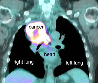
Lymph Nodes PET scan biopsy - Lung cancer - Inspire. The PET scan on the right lights up the structures that contain cancer thus helping prove that the cancer has already spread to these lymph nodes and changing the patients stage from stage I to stage III and eliminating surgery as a good treatment option. 1 days ago Sep 14 2019 I had no lymph nodes light up on my PET scan either.
Had bilatteral mascetomy with lymph node dissecetion on 12-17-12. Appears to be a single node. And with it being an 8mm nodule it may not even light up on the PET.
From the lower margin of the cricoid to the clavicles and the upper border of the manubrium. PET coronal image b show multiple mediastinal lymph nodes high FDG uptake arrow. I dont have an answer for you as this is all new for me as well and today when I met with the radiation oncologist I was told that I needed a PET scan before treatment can begin due tp enlarged lymph nodes from my CT scan.
If you are lower stage and going right into surgery lymph nodes will always be taken from different stations for testing.
![]()
Pet Ct Transaxial Images Showing Fdg Avid Right Hilar Lymph Node Nodal Download Scientific Diagram
Pet Imaging 101 Pet Scans

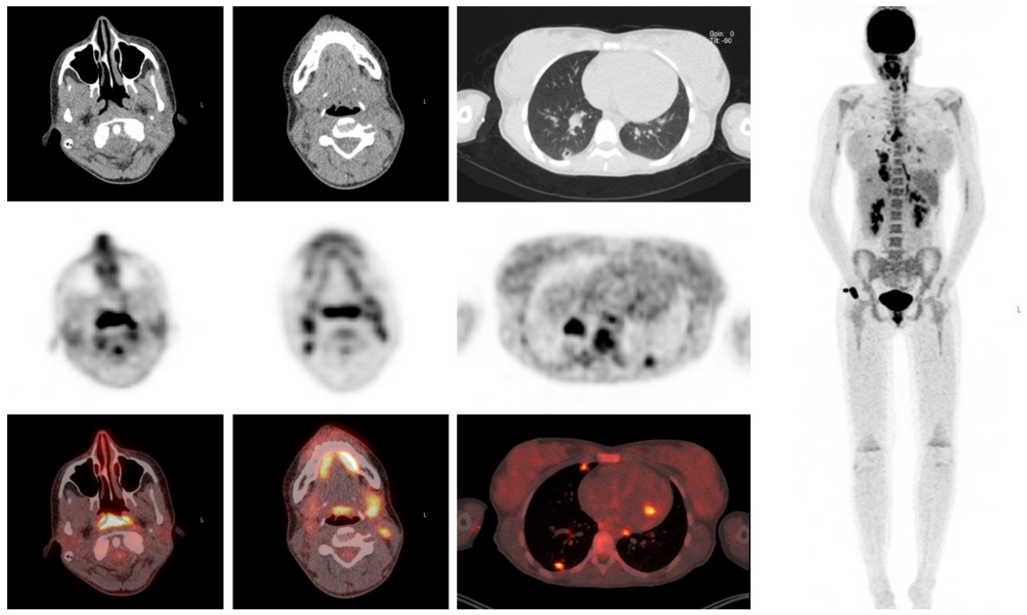
Figure1 18 F Fdg Pet Imaging Showing Hypermetabolic Lesions An Download Scientific Diagram

Fdg Pet Ct Scan Shows A Symmetrical Uptake In Fdg Positive Lymph Nodes Download Scientific Diagram
![]()
Pet Ct Transaxial Images Showing Fdg Avid Right Hilar Lymph Node Nodal Download Scientific Diagram
Pet Scans In Cancer Cases


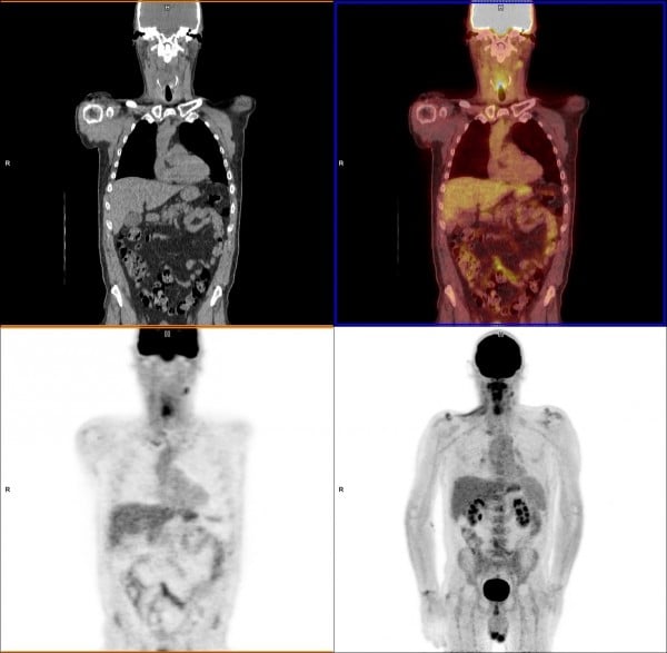
A Fdg Pet Ct Scan Demonstrated High Grade Metabolic Activity In The Download Scientific Diagram
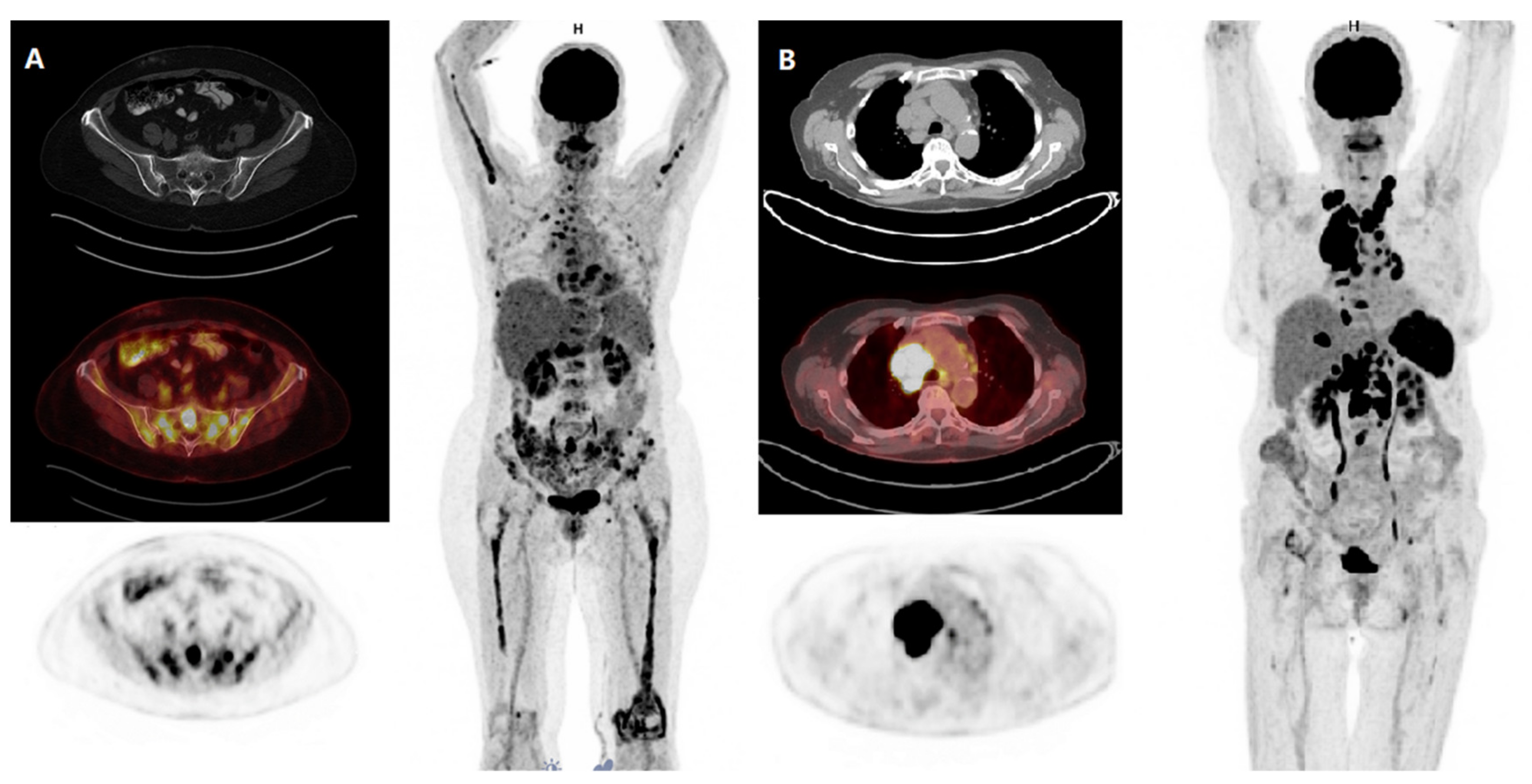
Cancers Free Full Text The Link Between Cytogenetics Genomics And Imaging Patterns Of Relapse And Progression In Patients With Relapsed Refractory Multiple Myeloma A Pilot Study Utilizing 18f Fdg Pet Ct Html
Pet Ct Open Air Mri Of Cen La

Pet Scans In Cancer Cases


Recommendations For Optimal Use Of Imaging Studies To Clinically Stage Mediastinal Lymph Nodes In Non Small Cell Lung Cancer Patients Lung Cancer
False Positive Fdg Pet Ct Findings In Enlarged Lymph Nodes In A Download Scientific Diagram
Pet Imaging 101 Pet Scans

Stage N3 Lymph Nodes A Axial Pet Ct Image Of The Chest Shows A Download Scientific Diagram

N Staging In Large Cell Neuroendocrine Carcinoma Of The Lung Diagnostic Value Of 18f Fdg Pet Ct Compared To The Histopathology Reference Standard Springerlink
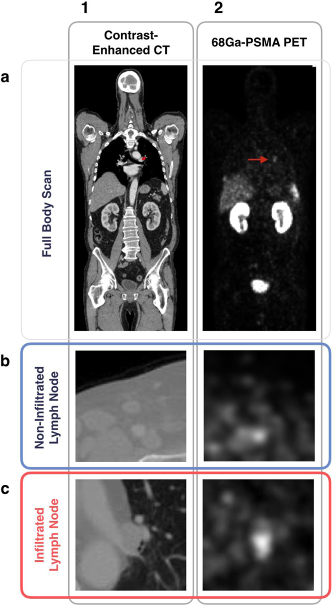
Prostate Cancer Nodal Staging Using Deep Learning To Predict 68ga Psma Positivity From Ct Imaging Alone Scientific Reports
Pet Scans In Cancer Cases
Pet Scans In Cancer Cases

Pet Scans In Cancer Cases

Post a Comment for "Mediastinal Lymph Nodes Light Up On Pet Scan"